Radiology is that amazing branch of medicine that gives doctors a superpower to see inside the human body without the need for surgery. By using advanced imaging techniques, the specialty of radiology transforms the invisible into clear and detailed images, revealing the secrets of diseases, injuries, and complex body anatomy. The discovery of radiology and its medical uses has caused a real revolution in the world of diagnosis and treatment, and today it has become an indispensable cornerstone in every modern hospital and clinic. Every radiographic image is a story told by the body, and the radiologist reads and interprets it to provide the treating physician with an accurate map that guides them toward the correct diagnosis and optimal treatment plan. The role of radiology is no longer limited to diagnosis, but has evolved to include guided and precise treatment through what is known as interventional radiology.
This comprehensive and integrated article will delve into the world of medical radiology, where we will explore its history and pivotal role, learn in detail about its different types and wide uses in detecting diseases, and highlight its many benefits. We will also address how examinations are performed and their results are analyzed, and provide a look at the future of this specialty that continues to push the boundaries of medical vision.
What is the Specialty of Radiology?
To clarify the nature of this important medical field, the specialty of radiology is the science that uses medical imaging techniques to diagnose and treat diseases. This specialty includes a wide range of methods that produce detailed images of internal organs, bone structures, and soft tissues, making radiology an indispensable tool in modern medical practice:
- The science of diagnostic imaging: This science is known as diagnostic imaging, as it relies on interpreting medical images to reach an accurate diagnosis of medical conditions.
- A medical specialty: A radiologist is a doctor who has completed specialized training after medical school, focusing on the use and interpretation of different imaging techniques.
- A dual role: The role of “radiology” is not limited to diagnosis only (diagnostic radiology), but extends to include performing guided and precise treatments using imaging techniques (interventional radiology).
- Diverse techniques: The specialty of radiology includes a variety of techniques, each with its own uses and physical principles, such as X-rays, ultrasound, and magnetic resonance imaging.
The Role of Radiology in Modern Medicine
The specialty of radiology plays a central and crucial role in almost every aspect of healthcare. From the emergency center to operating rooms, and from oncology clinics to pregnancy follow-up, important medical decisions now heavily rely on the accurate information provided by radiology examinations, which highlights its importance as an indispensable tool:
- Accurate and rapid diagnosis: Radiology provides the ability to determine the nature, location, and extent of diseases with great accuracy, which allows doctors to start the appropriate treatment quickly, which is crucial in cases such as strokes and cancer.
- Guiding treatment decisions: Radiology images help determine whether a patient needs surgery or can be treated with medications, and they are also used to accurately plan surgical procedures before they are performed.
- Monitoring treatment response: Radiology examinations are used periodically to monitor the response of tumors to chemotherapy or radiation therapy, evaluate the healing of fractures, and follow the progression of chronic diseases.
- Early detection and prevention: Some types of radiology, such as mammography, play a vital role in screening programs for the early detection of diseases before symptoms appear, which significantly increases the chances of recovery.
- Reducing the need for exploratory surgery: Before the advent of advanced radiology, doctors sometimes had to perform surgeries just to know what was inside the body. Now, radiology provides this information in a non-surgical way.
What are the Types of Radiology?
The field of radiology includes a variety of imaging techniques, each of which uses a different physical principle to produce images and has its own advantages and uses. Understanding these different types helps to realize the extent and development of the specialty of radiology:
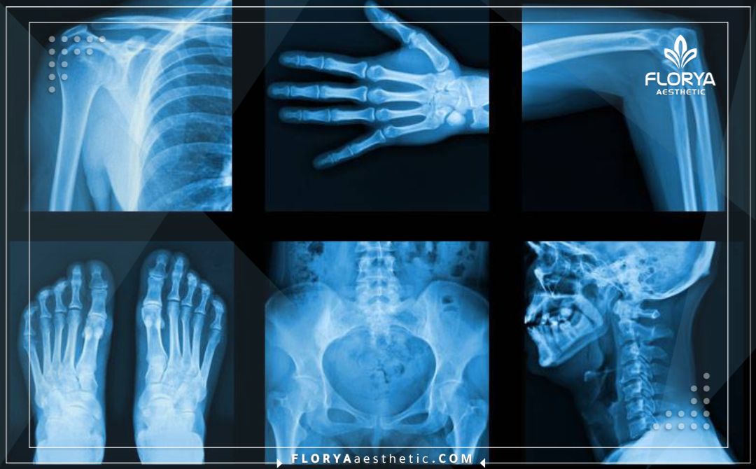
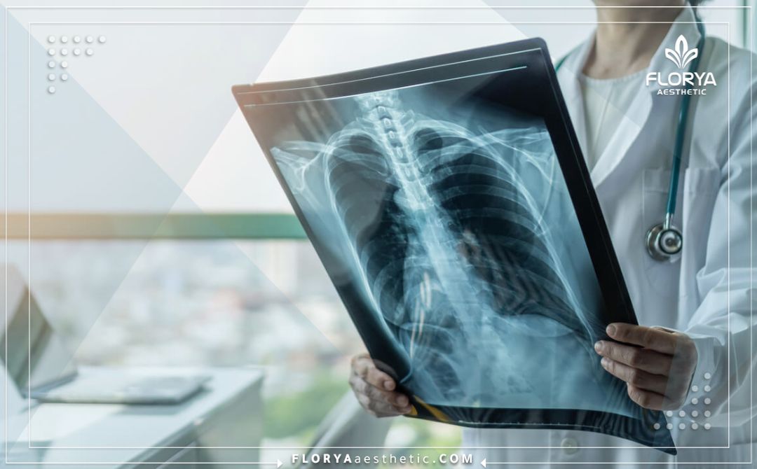
- X-ray: It is the oldest and most famous type of radiology. It uses a small beam of ionizing radiation to produce images of bones and internal structures, where bones appear white because they absorb most of the radiation.
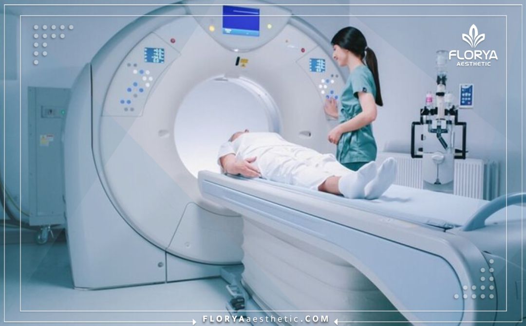
- Computed Tomography (CT Scan): An advanced form of X-ray, where the X-ray machine rotates around the body and takes a series of images from different angles. The computer combines these images to create detailed, three-dimensional cross-sections.
- Magnetic Resonance Imaging (MRI): It does not use ionizing radiation in some parts of the body. Instead, it uses a powerful magnetic field and radio waves to produce high-resolution images of soft tissues such as the brain, muscles, ligaments, and joints.
- Ultrasound: It uses high-frequency sound waves to create live images of internal organs. It is completely safe and does not use any radiation, which makes it ideal for pregnancy follow-up and examining abdominal organs.
- Nuclear Medicine / PET Scan: It involves injecting a very tiny amount of a radioactive substance into the body. A special camera tracks this substance and how it is distributed in the organs, which provides information about the function of the organ, not just its structure.
How to Perform an Examination Using Radiology and Analyze the Results
The process of performing a radiology examination includes organized steps that begin with preparation and end with a precise analysis of the images by a specialist doctor. Understanding this process helps to reduce patient anxiety and clarifies the specific role played by the radiology team:
- Preparation for the examination: The instructions depend on the type of examination. Some CT or nuclear radiology examinations may require fasting for a few hours or drinking a contrast fluid (dye) to improve the clarity of the images.
- Performing the examination: First, the radiology technician places the patient in the correct position on the examination table. The machine is turned on to take the images, and the patient may be asked to hold their breath for a few moments to prevent image blurring due to movement. Most examinations take between 15 to 60 minutes.
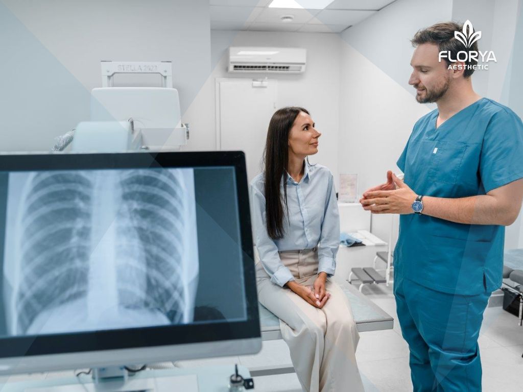
- Analyzing the results: After the images are obtained, they are sent electronically to the radiologist. The radiologist, a doctor specializing in interpreting medical images, examines the images with extreme care, and compares them to previous images if they exist. Then the radiologist writes a detailed report describing what he sees and provides his diagnosis or diagnostic impression, and then sends this report to the treating physician who requested it.
The Wide Medical Uses of Radiology in Detecting Diseases
The specialty of radiology is used to diagnose and monitor a huge range of diseases and injuries that affect all body systems. The versatility of radiology techniques makes it a powerful and indispensable diagnostic tool in daily medical practice:
- In bone and joint diseases: X-rays are primarily used to diagnose fractures, joint dislocations, arthritis, and to evaluate bone alignment after surgery.
- In nervous system diseases: Magnetic resonance imaging (MRI) is the best for diagnosing brain tumors, strokes, multiple sclerosis, spinal cord injuries, and herniated discs.
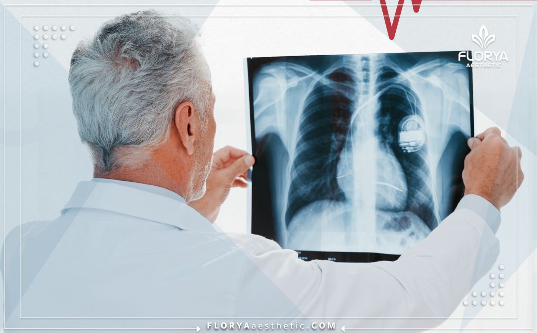
- In chest and heart diseases: Chest X-rays are used to evaluate the lungs and heart and detect pneumonia. CT scans are used to evaluate blood vessels in the lung and heart.
- In abdominal and digestive system diseases: Ultrasound and CT scans are used to examine the liver, gallbladder, pancreas, and kidneys and detect stones, tumors, and infections.
- In oncology (cancer): The specialty of radiology plays a crucial role in detecting tumors, determining their size and location, finding out if they have spread (tumor staging), guiding biopsies, and monitoring the response to treatment.
- In women’s and children’s health: Mammograms are used for the early detection of breast cancer. Ultrasound is used to monitor the growth and health of the fetus during pregnancy.
Treating Diseases Using Radiology
Radiology is no longer limited to detection, but has become an effective therapeutic tool in many medical specialties. This technique relies on precisely guiding specific doses of radiation to certain areas of the body with the aim of eliminating diseased cells or stopping their growth, while minimizing damage to the surrounding healthy tissues. Doctors use radiology as a primary or supplementary treatment in addition to surgery or medications, depending on the patient’s condition. Among the most prominent diseases that benefit from radiology treatment and limiting its effect are:
- Treatment of all types of cancerous tumors.
- Treatment of some thyroid diseases.
- Bone and joint diseases.
- Cases of benign prostatic hyperplasia.
- Small benign tumors.
Benefits and Advantages of Using Radiology
The use of radiology in medicine offers tremendous benefits to both patients and doctors, and these benefits have led to a significant improvement in diagnostic accuracy and a reduction in the need for painful and dangerous procedures. The progress in the field of radiology is directly reflected in the improvement of the quality of healthcare:
- Non-surgical and painless: Most radiology examinations are non-surgical procedures and do not cause any pain to the patient.
- High accuracy: Techniques such as MRI and CT scans provide extremely detailed images that allow for an accurate diagnosis of conditions that were difficult to detect in the past.
- Speed of results: In emergency cases, radiology examinations can be performed and results obtained within minutes, which helps in making quick and often life-saving treatment decisions.
- Guided treatment (Interventional Radiology): Interventional radiology allows doctors to perform complex treatments through very small incisions using image guidance. These procedures include opening blocked arteries, treating tumors, and taking biopsies, which reduces risks and speeds up patient recovery compared to traditional surgery.

Risks of Radiology to the Body
Despite the great benefits that radiology offers in diagnosis and treatment, excessive use or exposure to it without controls may cause health damage. This is because radiology relies on waves or particles that are able to penetrate body tissues, which may affect cells if medical safety rules are not followed. Hence comes the importance of using radiology in calculated doses, under specialized medical supervision, while providing protection means for patients and the medical team. Among the most prominent potential risks of radiology to the body are:
- Increased likelihood of damage to healthy cells.
- Exposure to problems in the bone marrow at high doses.
- Temporary disturbances in the reproductive system.
- The possibility of rare skin burns.
- Cumulative effects that may increase the risk of some chronic diseases. Thus, we find that radiology is a double-edged sword; it is a lifesaver when used correctly, and it can be dangerous if handled improperly.
Costs of Using Radiology in Detecting and Treating Diseases
The costs of radiology services are a large part of healthcare expenses, and they vary greatly based on the complexity of the technique used, the part to be imaged, and the geographical location. The following table shows the estimated costs of some common examinations and procedures in the field of radiology:
| Examination / Procedure | Description | Average Cost (in USD) | Notes |
| X-ray | A simple image of one area (such as the chest or hand). | $100 – $400 | It is considered the least expensive of all types of radiology examinations. |
| CT Scan | A sectional image of one area (such as the head or abdomen) without dye. | $800 – $3,000 | The cost increases by about 30-50% when using intravenous dye. |
| MRI | An MRI image of one area (such as the knee or brain) without dye. | $1,200 – $4,500 | It is considered one of the most expensive diagnostic examinations due to the cost of the device and the complexity of the technique. |
| Ultrasound | An examination of one area (such as the abdomen or pelvis). | $300 – $1,000 | Its cost is reasonable and it is completely safe. |
| PET Scan | A full-body examination often used in oncology. | $3,000 – $8,000 | A high-cost examination used to evaluate tissue functions and the spread of cancer. |
| Interventional Radiology (such as angioplasty) | A therapeutic procedure to open a blocked artery. | $15,000 – $50,000+ | The cost includes the procedure, the materials used, and the hospital stay. |
Prices of Using Radiology in Detecting and Treating Diseases
The prices of radiology services vary greatly according to the previous factors. Here is a list of average prices for some common examinations that are considered essential in medical diagnosis:
- Chest X-ray: Usually ranges from $100 – $350 USD.
- Mammogram: Ranges from $150 – $500 USD.
- Head CT scan without dye: Ranges from $700 – $2,000 USD.
- Lumbar spine MRI: The cost ranges from $1,500 – $4,000 USD.
- Pregnancy ultrasound: Ranges from $200 – $600 USD.
- Bone density scan (DEXA Scan): The cost ranges from $150 – $400 USD.
The Best Radiologists in the World
The field of radiology includes global leaders who have contributed to the development of imaging techniques and their clinical applications, which has led to a huge improvement in diagnostic accuracy. Here are five of the prominent experts in this field:
- Dr. Hedvig R. Hricak: Location: Department of Radiology, Memorial Sloan Kettering Cancer Center, New York, USA. About: A global pioneer in the field of tumor imaging, especially using MRI to diagnose and monitor prostate cancer and gynecological diseases. Her research has contributed to setting international standards in oncology radiology.
- Professor Gabriel P. Krestin: Location: Department of Radiology and Nuclear Medicine, Erasmus University Medical Center, Rotterdam, Netherlands. About: A prominent European expert and former president of the European Society of Radiology. He has great contributions to the development of MRI, CT scans, and the applications of artificial intelligence in radiology.
- Dr. Jeffrey S. Ross: Location: Mayo Clinic, Arizona, USA. About: One of the most prominent neuroradiology experts in the world. He is known for his deep expertise in interpreting complex MRI images of the brain and spine, and he is the author of many reference books in this field.
- Professor Sir Michael Brady: Location: Department of Oncology Sciences, University of Oxford, UK. About: A leading scientist and researcher in the field of medical image analysis. Although his background is in engineering, his work in developing artificial intelligence algorithms to analyze radiology images (especially mammograms) has a huge impact on the future of the specialty.
- Dr. Sanjiv Sam Gambhir: Location: Head of the Department of Radiology at Stanford University School of Medicine, California, USA. About: He is considered one of the greatest and oldest pioneers of molecular imaging and nuclear medicine. His pioneering work in developing early cancer detection techniques using radiology still influences the field today.
The Best Medical Centers Specialized in Radiology
Some global centers and hospitals are distinguished by having the latest radiology devices and the best experts, which makes them reference centers for accurate diagnosis and advanced treatments. Here are five of these leading centers in the world:
- Mayo Clinic – Department of Radiology: Location: Rochester, Minnesota, USA. About: It is consistently ranked as one of the best radiology departments in the world. It is known for its collaborative approach, where radiologists work closely with doctors from all specialties to provide an integrated diagnosis.
- Massachusetts General Hospital – Department of Radiology: Location: Boston, Massachusetts, USA. About: The main teaching hospital for Harvard Medical School. It is known for being a leading center in research and innovation, especially in the field of neuroradiology and functional imaging.
- University of California, San Francisco (UCSF) – Department of Radiology and Biomedical Imaging: Location: San Francisco, California, USA. About: A leading center in the development and application of advanced imaging techniques, especially in MRI and molecular imaging.
- Memorial Sloan Kettering Cancer Center – Department of Radiology: Location: New York, USA. About: One of the best cancer centers in the world, and its radiology department is fully specialized in tumor imaging, which provides unparalleled expertise in diagnosing and monitoring cancer using the latest techniques.
- Heidelberg University Hospital – Department of Diagnostic and Interventional Radiology: Location: Heidelberg, Germany. About: One of the most prominent European centers in the field of radiology, and it has a global reputation in the field of interventional radiology and tumor imaging, and it is known for its adoption of the latest digital and artificial intelligence technologies.
Frequently Asked Questions
Are radiology examinations safe? And is there a risk from radiation?
Most radiology examinations are very safe. Techniques such as MRI and ultrasound do not use any ionizing radiation. As for X-rays and CT scans, they use very low doses of radiation, and the great diagnostic benefit is always balanced with the very small risks, with care taken to use the lowest possible dose.
What is the difference between a radiologist and a radiology technician?
A radiology technician is the person who is technically qualified to operate imaging devices and perform the examination for the patient. As for a radiologist, he is a specialist doctor who has completed full medical training and then specialized in reading and interpreting these medical images and writing diagnostic reports.
What is “dye” or “contrast material” and why is it used?
A contrast material (dye) is a A special fluid that is given to the patient (orally or by intravenous injection) before some radiology examinations (especially CT scans and MRIs). This material helps to highlight certain organs or blood vessels and make them clearer in the images, which leads to a more accurate diagnosis.
Why don’t I get my results directly from the radiology technician?
Because the radiology technician is trained to take high-quality images, but is not medically qualified to interpret them. The radiologist (the doctor) must carefully review the images and write an official report, and this process requires time and expertise to ensure accuracy and avoid any misdiagnosis.
Conclusion
In conclusion of this journey into the world of radiology, it becomes clear that this specialty is truly the sharp eye of modern medicine. The ability to see inside the body without surgical intervention has transformed the way we understand, diagnose, and treat diseases. From a simple X-ray that reveals a fracture, to a complex PET/CT scan that maps the spread of cancer, the specialty of radiology plays an indispensable role in every step of the patient’s journey. With the amazing developments in artificial intelligence, molecular imaging, and interventional radiology, the future of radiology promises to be more accurate, faster, and safer, which opens new horizons for early detection and personalized treatment. The specialty of radiology will always be the compass that guides medicine, lights the way for doctors, and offers hope to patients all over the world.
- American College of Radiology (ACR). Clinical Tools & Reference. Available at: https://www.acr.org/Clinical-Resources/Clinical-Tools-and-Reference
- RadiologyInfo.org. Radiology Information Resource for Patients. Available at: https://www.radiologyinfo.org/en/
- Radiopaedia.org. Radiopaedia – A Collaborative Radiology Resource. Available at: https://radiopaedia.org/
- Radiology Assistant (Netherlands). Radiology Assistant Educational Site. Available at: https://radiologyassistant.nl/
- Society of Interventional Radiology (SIR). Society of Interventional Radiology. Available at: https://www.sirweb.org/